What Is a Mammogram? Understanding the Utility and Purpose
October 12, 2023Categories: Breast Cancer

If you are nearing your 40s, you may have heard about the importance of scheduling regular mammograms to screen for breast cancer. Most women should start these screenings at age 40. Women with a family history or other risk factors for breast cancer may need to start them earlier. Even if you know you need one, you might be wondering what a mammogram is and how it can help you.
“Women probably understand, on some level, what a mammogram is — a test that detects breast cancer early, when it’s easier to treat,” says Amy Hane, BSN, RN at the Beaufort Memorial Breast Health Center. “But they may have a lot of questions about how the test is done and what we’re looking for.”
Read More: What to Expect During Your First Mammogram
Screenings for Prevention Versus Diagnosis
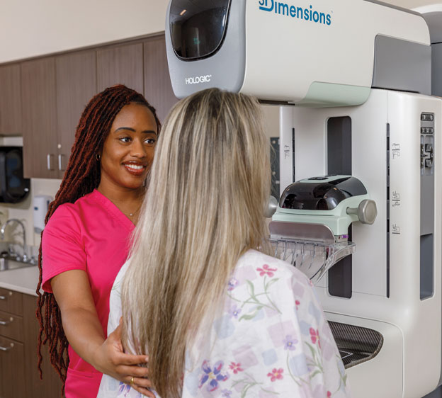
The mammograms we encourage women to get each year are screening mammograms. During these exams, a mammographer takes X-ray images of each breast to look for abnormalities and other signs that you may have breast cancer or another breast condition. “The mammographer is the first set of eyes for the radiologist,” Hane says. “While the radiologist is ultimately responsible for reading the scan, astute mammographers have the first set of eyes on the images and can anticipate what the radiologist may need.”
A radiologist is a medical doctor who uses diagnostic techniques such as mammography, ultrasound, breast MRI and minimally invasive biopsy to diagnose cancer and other diseases. The radiologist’s interpretation of the imaging findings is billed separately from the procedure, so you should expect to receive two invoices following your screening mammogram appointment.
After the mammogram, the radiologist reviews the images and looks for noticeable changes in your breasts from your previous screening (if you’ve had one before) and abnormal tissue that might need further investigation.
If something unusual appears, the radiologist will use a standing order (written protocols that authorize designated members of the health care team to complete certain clinical tasks without having to first obtain a physician order) or call the referring physician for an order to perform a diagnostic mammogram. These mammograms involve X-ray images, just like a screening mammogram, but the mammographer takes more images, so the exams typically last longer.
Read More: Screening vs. Diagnostic Mammogram: What’s the Difference?
The Power of 3D Screening Technologies
For many years, mammography X-rays were 2D images. 3D mammograms, also known as tomosynthesis, have become more common because they take clearer and more detailed photos of breast tissue than 2D mammograms. Beaufort Memorial began using the 3D technology more than 10 years ago and in early 2023 began using the 3D technology as the standard of care for all patients receiving screening mammograms.
The improved image quality includes many advantages over conventional 2D images, including:
- Fewer call-backs. Since radiologists can see a detailed view of your breast tissue from just one screening, you have less of a chance of being called back for additional tests.
- A 40% increase in breast cancer detection. 3D imaging technology has brought with it a high success rate in detecting breast cancer before symptoms start showing.
- 15% fewer false-positive results. Your test results are false-positive if they suggest you have a condition you don’t actually have. 3D screening technologies can help radiologists avoid this problem.
Many insurance providers now cover 3D mammography for screening mammograms, but if you aren’t sure, call your provider.
Read More: Answers to 5 Questions About Mammograms
What About Dense Breast Tissue?
Around half of all women over 40 have dense breasts. Breast density refers to the amount of fibrous and glandular tissue your breasts have. Because dense tissue can look white or gray in X-ray images, your radiologist might have a difficult time differentiating it from cancer. Women with dense breasts may need additional screenings, such as automated breast ultrasound screening (or ABUS).
Read More: Breast Cancer Screening for Women With Dense Breasts
The ABUS technology uses sound waves to identify potentially cancerous tissue in dense breasts, which may be missed by mammography. When radiologists use both mammogram and ultrasound technology, there is an overall increase in breast cancer detection.
Is it time for your annual screening mammogram? Call 843-522-5015 today or submit a request to schedule it at one of our three convenient locations in Beaufort, Hilton Head or Okatie.

